
Why Normal Imaging Does Not Mean Nothing Is Wrong
Your MRI Is Normal. So Why Do You Still Feel Like This?
You got the imaging done. You waited for the results. And then someone told you everything looks normal.
And you sat there thinking: then why does it still hurt?
This is one of the most common experiences people describe when they first come to Spine Pain and Performance Center. Not dramatic injury findings. Not clear-cut pathology. Just a normal scan and a body that still does not feel right.
What most people do not know is that this is not a contradiction. It is actually one of the most predictable patterns in musculoskeletal health. Normal imaging and abnormal mechanics happen together all the time. And understanding why changes everything about how you approach the problem.
What Imaging Is Actually Designed to Find
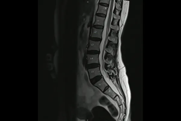
Diagnostic imaging, whether MRI, CT, or standard X-ray, is extraordinarily good at what it was built to do. It finds tissue damage. Disc herniations. Fractures. Tumors. Structural pathology that requires medical intervention.
For those things, imaging is essential. It is the right tool.
But here is what it was never designed to capture: the way your body moves, loads, and compensates as a living system. A still image of your spine tells you what the architecture looks like in a lying-down or standing position at one moment in time. It does not tell you how that architecture behaves under the demands of your actual life.
It does not show where a spinal segment has lost its normal motion. It does not reveal how a restriction in one area has shifted load to the joints above and below it over months or years. It does not capture the compensation pattern your nervous system has quietly built around a subluxation that has been active far longer than the pain has.
This is not a flaw in imaging. It is just the limit of the tool. The problem comes when that limit is not communicated clearly, and a normal result gets interpreted as if nothing is wrong.
The Body Compensates Before It Breaks Down
This is one of the most important things I want patients to understand, because it reframes what their experience actually means.
The body does not go from healthy to symptomatic overnight. There is a long middle phase where compensation is happening silently. A segment becomes restricted. The nervous system routes around it. Adjacent joints absorb the excess load. Muscles alter their recruitment patterns to protect the area. Posture shifts gradually.
All of this happens before pain becomes consistent or severe. The body is brilliant at adapting. It will work around a problem for a very long time before you feel it clearly.
By the time the pain is consistent enough to send someone for imaging, the compensation has often been active for years. And that compensation, not the original restriction, is usually what is generating the symptoms.
Research supports this consistently. A landmark study published in the New England Journal of Medicine found that a significant percentage of people with no back pain at all showed disc bulges, protrusions, and degeneration on MRI. A 2015 systematic review in the American Journal of Neuroradiology confirmed that imaging findings commonly considered abnormal are present in large proportions of pain-free individuals across all age groups. And a 2019 study in the Journal of Orthopaedic and Sports Physical Therapy documented that movement dysfunction and altered motor control patterns are poor predictors of imaging findings in chronic low back pain populations.
The scan is not lying. The scan just cannot see what the body has been doing to manage the problem.
What a Structural X-Ray Assessment Reveals That Diagnostic Imaging Does Not
This is where the Gonstead approach offers something genuinely different, and it is worth explaining clearly.
Gonstead chiropractic uses full-spine, weight-bearing X-ray analysis as part of its assessment. This is not the same type of imaging ordered for pathology screening. The purpose, the positioning, and the interpretation are different.
A Gonstead structural X-ray is taken with you standing, under the load of your own body weight, in the posture your spine actually holds day to day. It is a full-spine view that allows us to see how your spinal architecture is distributing that load from the base up.
We are looking at disc space height and how it changes from one level to the next. We are looking at vertebral alignment and rotation. We are looking at the angle of the pelvis and how it influences the segments above. We are looking at the cumulative effect of compensation patterns that have developed over time.
When this is combined with instrumentation to detect thermal asymmetry along the spine, motion and static palpation to identify where segments have lost joint play, and a full postural evaluation, we build a mechanical picture that tells us something a standard MRI cannot.
We can see where the compensation chain started. We can identify the primary subluxation that has been driving the pattern. And we can understand why the painful area hurts without that area necessarily being the source.
The "Normal Scan, Ongoing Pain" Patient
If I had to describe the person this piece is written for, it is someone who has done everything they were supposed to do. They got the imaging. They followed the recommendations. They may have done physical therapy. They were told to rest or strengthen or stretch.
And they still do not feel right. Not dramatically broken. Not in crisis. Just a persistent background of something that flares, something that limits, something that has not resolved despite everything they have tried.
That person is not imagining anything. They are not exaggerating. They have a compensation pattern that has never been properly mapped and a mechanical driver that has never been precisely identified.
The imaging was appropriate. It just answered a different question than the one they needed answered.
What Actually Needs to Be Evaluated
If normal imaging has not explained your pain, the next question is not which specialist to see next or whether something more sinister was missed. In most cases of recurring mechanical pain, the next question is: has anyone evaluated how your whole spine is moving and loading as a system?
Not just the area that hurts. The whole spine. Underweight. In the position it holds all day.
Because when you assess the system, you often find the answer that the symptom-focused search kept missing. Not because something dangerous was overlooked. Because the right mechanical question was never asked.
Key Takeaways
Normal imaging does not mean nothing is wrong. It means no tissue damage requiring medical intervention was found.
Diagnostic imaging cannot show compensation patterns, subluxations, or how the spine distributes load under real-world conditions.
The body compensates silently for a long time before pain becomes consistent. By the time you scan it, the compensation is often well established.
Gonstead structural X-ray analysis is taken weight-bearing, full-spine, and interpreted for mechanical load distribution, not pathology screening.
Normal imaging with ongoing pain is not a dead end. It is a signal that the right assessment has not happened yet.
If you have been told your imaging is normal and you are still dealing with recurring or persistent pain, the conversation does not have to end there. A full Gonstead structural assessment gives you a mechanical answer that imaging was never designed to provide.
That answer is where the path forward actually begins.
Explore the Movement Intelligence Assessment at Spine Pain and Performance Center.
References
1. Jensen MC et al. Magnetic resonance imaging of the lumbar spine in people without back pain. N Engl J Med. 1994. https://pubmed.ncbi.nlm.nih.gov/8022489/
2. Brinjikji W et al. Systematic literature review of imaging features of spinal degeneration in asymptomatic populations. AJNR Am J Neuroradiol. 2015. https://pubmed.ncbi.nlm.nih.gov/25430861/
3. Laird RA et al. Comparing lumbar spinal kinematics in people with and without back pain. J Orthop Sports Phys Ther. 2019. https://pubmed.ncbi.nlm.nih.gov/25627148/
4. Jarvik JG, Deyo RA. Diagnostic evaluation of low back pain with emphasis on imaging. Ann Intern Med. 2002. https://pubmed.ncbi.nlm.nih.gov/12353946/